Ascaris lumbricoides: Ascaris lumbricoides is a large parasitic roundworm that commonly infects humans. It’s one of the most prevalent parasitic infections globally, affecting an estimated 807 million to 1.2 billion people.
Ascaris lumbricoides
Classification:
Phylum …. Aschelminthes
Class …. Nematoda
Order …. Ascaroidea
Genus …. Ascaris
Species …. lumbricoides
Ascaris lumbricoides is an endoparasite in the small intestine of man lying freely in the lumen. It has existed in humankind since the beginning of time. It is cosmopolitan in distribution. It is found more commonly in children than in adults. Sometimes it migrates from intestine to stomach and comes out through the mouth or nostrils of the host.
As many as 1000 to 5000 adult worms may inhabit a single host. Mode of nutrition is holozoic, as it feeds on host’s partly digested food by sucking action of its pharynx. It produces anti-enzymes to protect itself from the action of the host enzyme. Sexual dimorphism is well distinct; only sexual reproduction takes place, asexual reproduction does not occur. Life cycle is simple and monogenetic.
External features
Shape: Ascaris lumbricoides is elongated, cylindrical, and tapering at both ends. It is a large sized nematode showing sexual dimorphism, i.e., sexes are separate.
Size: The female is 20–41 cm (8–16 inches) long and 4–6 mm in diameter, but the male is smaller, being 15–31 cm (6–12 inches) and 2–4 mm in diameter; its posterior end is curved ventrally.
Colour: Generally nematodes have no colour, the external cuticle is whitish or yellowish but some, like Ascaris have a definite reddish tint caused by the presence of haemoglobin.
Morphology: The anterior end of both the sexes exhibits the same structure. The body is covered with a smooth, tough and elastic cuticle which is striated transversely and gives the pseudosegmented appearance to the worm.
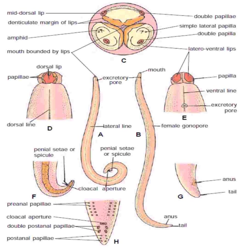
The cylindrical body has four longitudinal epidermal chords visible externally, the narrow one mid-dorsal, one mid-ventral and two thick ones are lateral. The dorsal and ventral chords appear white, while the laterals appear brown.

D—Anterior end in dorsal view ; E—Anterior end in ventral view ; F—Posterior end of
male ; G—Posterior end of female ; H—Posterior end of male in ventral view showing
papilla.
In nematodes the anterior mouth is bounded by six lips or labia but they are reduced by fusion to three in Ascaris, one elliptical mid-dorsal and two oval latero-ventral in position. Therefore, the mouth of Ascaris lumbricoides is a triradiate aperture. The dorsal lip has 2 double sensory papillae, and each latero-ventral lip has 1 double sensory papilla; these four papillae form an outer labial circle though most nematodes have 6 papillae in the outer labial circle.
Nematodes also have an inner labial circle of 6 papillae, but in Ascaris, as in most parasitic nematodes, the papillae of the inner labial circle are absent. The lateroventral lips have a lateral papilla each and cuticular excavation called amphid which is reduced in parasitic nematodes. Amphids are olfactory chemoreceptors. The lips have fine teeth. Behind the lips there is a pair of cervical papillae, one on each side close to the nerve ring. All papillae are sensory.
Near the posterior end is a transverse anus with thick lips, but the male has a cloaca from which two equal chitinous spicules or penial setae project. In the male near the cloaca there are cuticular elevations ventrally, about 50 pairs of pre-anal papillae and 5 pairs of post-anal papillae, these are concerned with copulation. There is a short post-anal tail which is straight in the female but sharply curved in the male. The female genital aperture called vulva or gonopore is on the ventral side, about one-third of the length from the anterior end. Behind the lips, there is an excretory pore mid-ventrally.
Life Cycle: The adult worms (Ascaris lumbricoides) live in the small intestine. Females can produce around 200,000 eggs per day, which are passed in the feces. These eggs become infective in soil and, when ingested, hatch into larvae that migrate through the body before settling back in the intestines2.
Symptoms: Many people with ascariasis have no symptoms. However, heavy infections can cause abdominal pain, malnutrition, and intestinal blockage. In severe cases, the worms can migrate to other parts of the body, causing complications like lung issues or bile duct obstruction3.
Transmission: The infection is typically spread through ingestion of contaminated soil or food. Poor sanitation and hygiene practices are major risk factors3


