

Collection and Selection of Specimens or Tissue for Whole Mount: The accurate and precise collection and selection of specimens or tissues are paramount in the field of microscopy and pathology. These initial steps significantly impact the subsequent analysis and interpretation of results. This article will delve into the critical aspects of specimen collection and selection for whole mount, smearing, and testing procedures.
Collection and Selection of Specimens or Tissue for Whole Mount
Types of Specimens
Before discussing collection and selection, it’s essential to understand the different types of specimens commonly used in microscopy and pathology:
Whole organisms: Small organisms like nematodes, rotifers, or daphnia.
Tissue sections: Excised or biopsied portions of organs or tissues.
Cells: Isolated cells from blood, bone marrow, or other sources.
Fluids: Body fluids like blood, urine, cerebrospinal fluid, or sputum.
Microorganisms: Bacteria, fungi, or parasites.
Specimen Collection
The method of collection depends on the type of specimen and the intended analysis. Key considerations include:
Sterility: For microbiological or tissue culture studies, strict aseptic techniques are crucial to prevent contamination.
Timing: The optimal time for collection depends on the biological process being studied. For example, blood samples may be collected at different times of the day to assess hormone levels.
Quantity: Sufficient material is necessary for accurate analysis, but excessive amounts can interfere with procedures.
Storage: Proper storage conditions (temperature, humidity, preservatives) are essential to maintain specimen integrity.
Specimen Selection
The selection of appropriate specimens is crucial for obtaining meaningful results. Key factors include:
Representativeness: The specimen should accurately reflect the condition or process being studied.
Size and shape: The specimen should be suitable for the intended technique (e.g., whole mount, smear).
Quality: The specimen should be free from damage, artifacts, or contaminants.
Ethical considerations: Specimen collection should adhere to ethical guidelines, especially when involving human or animal subjects.
Techniques: Whole Mount, Smearing, and Testing
Techniques for Whole Mount
Whole mount preparation is a technique used in microscopy to examine entire small organisms or thin layers of tissue. It involves several steps to preserve the specimen’s structure and create a suitable mount for observation.
Steps Involved in Whole Mount Preparation
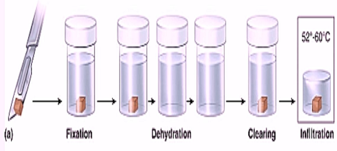
Fixation:
- This step preserves the specimen’s structure by killing and hardening the tissue.
- Commonly used fixatives include formalin, alcohol, and acetic acid.
- The choice of fixative depends on the type of tissue and the desired outcome.
Dehydration:
- Removes water from the tissue to prevent decay and improve embedding.
- Typically involves a graded series of alcohol solutions, from low to high concentrations.
- Other dehydrating agents like acetone or xylene can also be used.
Clearing:
- Replaces the dehydrating agent with a substance that has a refractive index similar to the mounting medium.
- Common clearing agents include xylene, toluene, and clove oil.
Embedding:
- Infiltrates the tissue with a solid medium for support and protection.
- Commonly used embedding media include paraffin wax, resin, or gelatin.
- The choice of embedding medium depends on the desired thickness of the final mount.
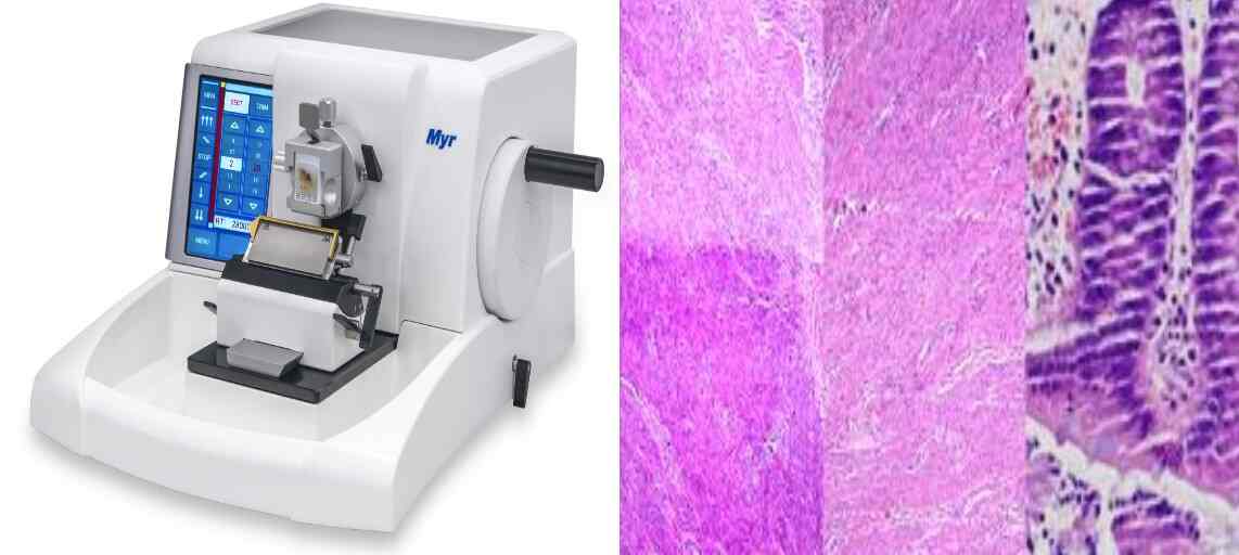
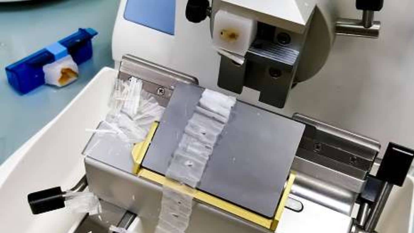
Sectioning:
- For larger specimens, sectioning may be necessary to create thin slices for observation.
- A microtome is used to cut the embedded tissue into sections.
Mounting:
- The sections or entire specimen are placed on a microscope slide and covered with a coverslip.
- A mounting medium with a refractive index similar to the clearing agent is used to prevent air bubbles and improve image clarity.
- Common mounting media include Canada balsam, DPX, and synthetic resins.
Specific Techniques
Whole mount of small organisms:
- Organisms like nematodes, rotifers, or daphnia can be directly mounted in a drop of water or glycerin on a slide.
- For long-term preservation, clearing and mounting in a resin-based medium is recommended.
Whole mount of tissue layers:
- Thin layers of tissue, such as skin or epithelium, can be prepared as whole mounts.
- Fixation, dehydration, clearing, and mounting steps are similar to those used for larger specimens.
Staining
To enhance contrast and visualize specific structures, staining techniques can be applied before or after mounting:
- Hematoxylin and eosin (H&E) stain: Commonly used for general tissue staining.
- Methylene blue: Stains nuclei and cytoplasmic granules.
- Gram stain: Differentiates bacteria based on cell wall structure.
- Periodic acid-Schiff (PAS) stain: Highlights carbohydrates and glycoproteins.
Techniques for Smearing
Smearing is a technique used in microscopy to create a thin layer of cells or microorganisms on a slide for examination. It’s a crucial step before staining and microscopic analysis.
Types of Smears
- Blood Smears: Used for examining blood cells and platelets.
- Bacterial Smears: For observing bacterial morphology and staining characteristics.
- Pap Smears: To detect abnormal cells in the cervix.
- Spinal Fluid Smears: For examining cells in cerebrospinal fluid.
General Smearing Technique
- Clean the slide: Ensure the slide is clean and grease-free for optimal results.
- Transfer the specimen: Using a sterile loop, pipette, or swab, transfer a small amount of the specimen to the center of the slide.
- Spread the specimen:
- Blood smear: Use a spreader slide to create a thin, even film.
- Bacterial smear: Spread the specimen in a circular motion to cover a small area.
- Other smears: The spreading technique may vary depending on the specimen type.
- Air dry: Allow the smear to air dry completely.
- Fixation: This step adheres the specimen to the slide and kills microorganisms. Common methods include heat fixation or methanol fixation.
Specific Smearing Techniques
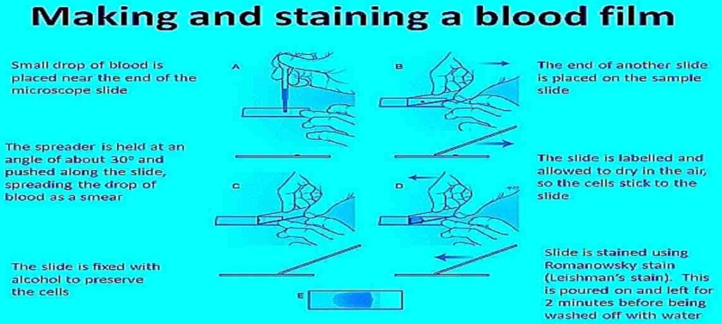
Blood Smear
- Wedge smear: The most common method, involving a spreader slide to create a feathered edge.
- Pull smear: Used for bone marrow aspirates.
Bacterial Smear
- From solid media: Emulsify a small amount of colony in a drop of water on the slide.
- From liquid media: Place a small drop of the culture on the slide.
Pap Smear
- Use a special spatula or brush to collect cells from the cervix.
- Transfer the cells to a slide and prepare a thin smear.
Testing: This encompasses a wide range of techniques, including biochemical, immunological, and molecular methods, to analyze specific components of the specimen.