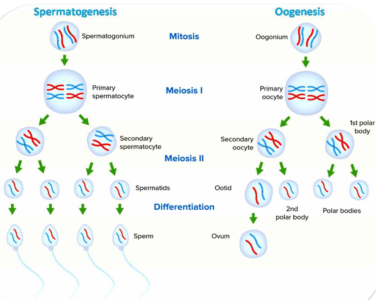
Gametes: Gametes are reproductive cells that contain a haploid number of chromosomes. This means that they have only one copy of each chromosome, as opposed to the diploid number of chromosomes found in other cells of the body. Gametes are produced in the gonads, which are the reproductive organs of the body. In males, the gonads are the testes, and in females, the gonads are the ovaries.
In addition to these basic features, some gametes also have other specialized structures. For example, sperm cells have a long tail that helps them to swim towards the egg. Egg cells, on the other hand, are usually larger and non-motile. They often have a protective layer around them to prevent them from being fertilized by multiple sperm cells.
Structure of Gametes:
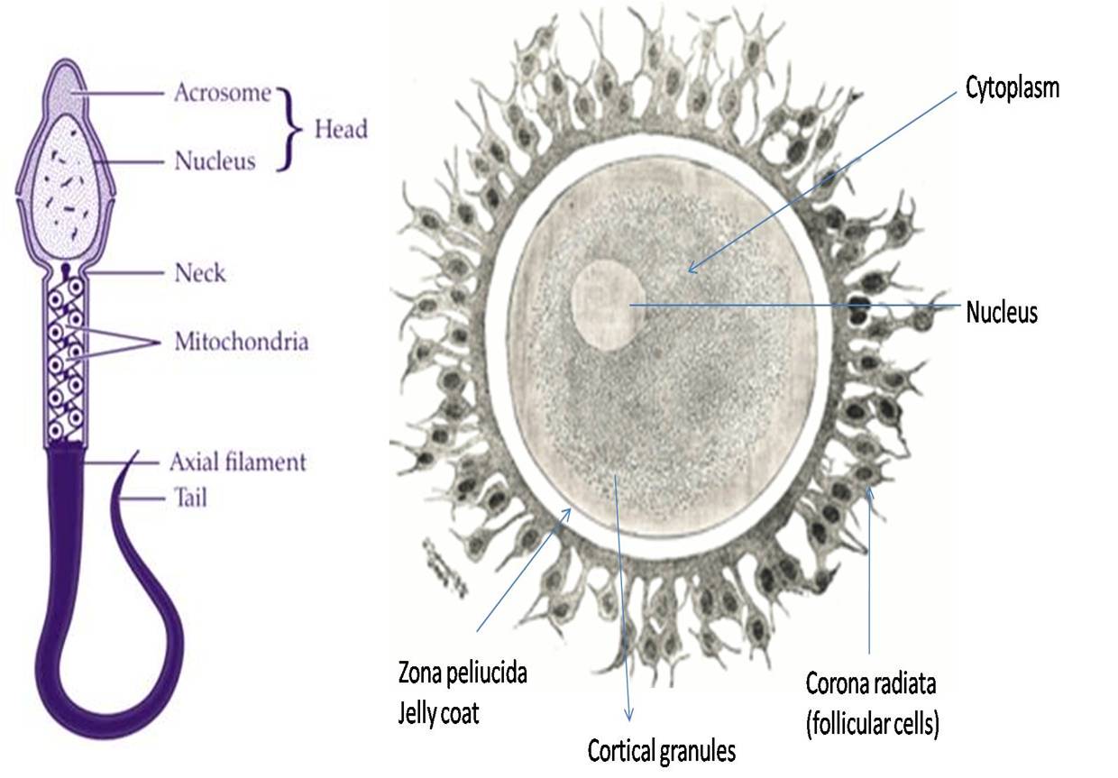
The structure of gametes varies depending on the species, but all gametes share some common features. First, they all contain a nucleus, which contains the genetic material of the cell. Second, they all have a plasma membrane, which is a thin layer that protects the cell and helps it to maintain its shape. The detailed structure of sperm and ovum as follows.
Structure of a Sperm
Sperm are the male gametes, or sex cells, in humans and other animals. They are produced in the testes and ejaculated during sexual intercourse. Sperm are microscopically small, measuring only about 60 micrometers in length. However, they are highly specialized cells that are adapted for the single purpose of fertilizing an egg.
Sperm have a distinctive structure that can be divided into three main parts: the head, the midpiece, and the tail.
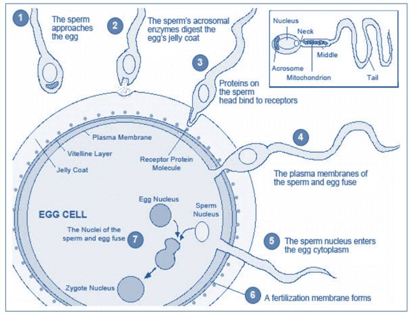
- Head: The head of the sperm contains the nucleus, which contains the genetic material of the father. The head is also covered by a cap-like structure called the acrosome, which contains enzymes that help the sperm to penetrate the egg.
- Midpiece: The midpiece of the sperm contains mitochondria, which are organelles that produce energy. The energy is used to power the tail of the sperm, which propels it through the female reproductive tract.
- Tail: The tail of the sperm is long and thin, and it is responsible for the sperm’s movement. The tail beats back and forth, like a propeller, to move the sperm towards the egg.
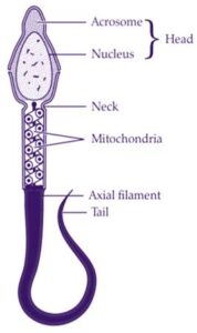
The entire sperm cell is covered by a plasma membrane, which is a thin layer that protects the cell and helps it to maintain its shape.
Function of the sperm structure
The structure of the sperm is specifically adapted to its function of fertilizing an egg. The head of the sperm contains the genetic material that will be passed on to the offspring. The midpiece contains the energy that the sperm needs to swim towards the egg. And the tail provides the sperm with the power to move through the female reproductive tract.
When a sperm cell encounters an egg, the acrosome releases enzymes that help the sperm to penetrate the egg’s protective layer. Once the sperm has entered the egg, the nucleus of the sperm fuses with the nucleus of the egg, forming a fertilized egg.
Structure of Ovum
The ovum, also known as an egg cell, is the female gamete in humans and other animals. It is produced in the ovaries and released during ovulation. The ovum is much larger than a sperm cell, measuring about 120 micrometers in diameter. It is also non-motile, meaning that it cannot move on its own.
The ovum has a complex structure that is essential for its role in reproduction. The main parts of the ovum are:
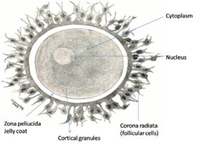
- Zona pellucida: The zona pellucida is a thick, protective layer that surrounds the ovum. It prevents multiple sperm cells from fertilizing the egg and helps to guide the sperm to the egg. It also contains proteins that bind to specific receptors on the sperm cell, helping to guide the sperm to the egg. Inside the zona pellucida is the cytoplasm of the egg cell. The cytoplasm contains all of the organelles and nutrients that the egg needs to develop into a new embryo. The nucleus of the egg cell is located in the center of the cytoplasm.
- Vitelline membrane: The vitelline membrane is a thin membrane that lies just beneath the zona pellucida. It helps to protect the ovum from damage.
- Cytoplasm: The cytoplasm is the jelly-like substance that fills the inside of the ovum. It contains all of the organelles and nutrients that the ovum needs to develop into a new embryo.
- Cortex: The cortex is a thin layer of cytoplasm that surrounds the ovum. It contains microvilli, which are small projections that help to transport nutrients into and out of the ovum.
- Nucleus: The nucleus is located in the center of the cytoplasm. It contains the genetic material of the ovum, which will be passed on to the offspring.
- Germinal vesicle: The germinal vesicle is the nucleus of the ovum. It is a large, round nucleus that contains the genetic material of the ovum.
- Nucleolus: The nucleolus is a small structure inside the germinal vesicle. It is responsible for producing ribosomes, which are organelles that synthesize proteins.
Vitellogenesis
Vitellogenesis is the process of yolk protein formation in the oocytes of non-mammalian vertebrates during sexual maturation. The term vitellogenesis comes from the Latin vitellus (“egg yolk”). Yolk proteins, such as Lipovitellin and Phosvitin, provide maturing oocytes with the metabolic energy required for development. Vitellogenins are the precursor proteins that lead to yolk protein accumulation in the oocyte. Vitellogenesis is a complex process that is regulated by a number of hormones, including estrogen, growth hormone, and insulin-like growth factor 1. Estrogen is the main hormone that stimulates vitellogenesis, and it is produced by the ovaries. Growth hormone and insulin-like growth factor 1 are produced by the liver and other tissues, and they help to promote the synthesis of yolk proteins.
The process of vitellogenesis begins with the synthesis of yolk proteins in the liver. The yolk proteins are then released into the bloodstream and transported to the developing oocytes. Once the yolk proteins reach the oocytes, they are taken up by the cells through receptor-mediated endocytosis. The yolk proteins are then stored in the cytoplasm of the oocytes, where they will be used to fuel the development of the embryo.
Vitellogenesis is an essential process for reproduction in non-mammalian vertebrates. Without vitellogenesis, the developing oocytes would not have the nutrients they need to develop into healthy embryos.
Steps involed in vitellogenesis:
- Estrogen is produced by the ovaries and stimulates the synthesis of yolk proteins in the liver.
- Yolk proteins are released into the bloodstream and transported to the developing oocytes.
- Yolk proteins are taken up by the oocytes through receptor-mediated endocytosis.
- Yolk proteins are stored in the cytoplasm of the oocytes, where they will be used to fuel the development of the embryo.
Vitellogenesis is a complex process that is regulated by a number of factors, including hormones, genes, and environmental conditions. It is essential for reproduction in non-mammalian vertebrates.
Fertilization of Gametes
When a sperm cell fertilizes an ovum, the two nuclei fuse together to form a new diploid nucleus. This new nucleus contains all of the genetic information that is needed to create a new organism.
The fertilized ovum cell then begins to develop into an embryo. The embryo grows and divides until it is ready to be born.
Conclusion
The structure of gametes is essential for their ability to reproduce. Sperm cells and egg cells have different structures, but they both contain the genetic material that is needed to create a new organism. The structure of the sperm is essential for its ability to fertilize an egg. The head, midpiece, and tail all play important roles in helping the sperm to reach the egg and deliver its genetic material. The structure of the ovum is essential for its role in reproduction. The ovum has a number of specialized structures that help to protect it, transport nutrients, and synthesize proteins. These structures are all necessary for the ovum to develop into a new embryo.

