Microtechnique: Microtechnique is a fascinating and essential field within biological sciences, providing invaluable insights into the microscopic structure and function of various specimens. This article delves into the definition, scope, and application of microtechnique, highlighting its significance and the advancements it brings to scientific research.
Definition of Microtechnique
Microtechnique refers to the collection of methods and procedures used to prepare and analyze specimens under a microscope. This field encompasses a variety of techniques aimed at enhancing the visualization and understanding of microscopic structures. These techniques are crucial for examining biological tissues, cells, and microorganisms, as well as materials science applications where the microscopic analysis of substances is required. This typically includes fixation, dehydration, embedding, sectioning, staining, and mounting.
Scope of Microtechnique
Microtechnique encompasses a broad spectrum of techniques applicable to various fields of study, primarily in the life sciences. The scope of microtechnique includes:
Botanical Microtechnique: Focuses on preparing plant tissues for microscopic examination.
Zoological Microtechnique: Deals with preparing animal tissues for microscopic study.
Microbiological Microtechnique: Involves preparing microorganisms for observation.
Histological Microtechnique: Specifically concerned with the preparation of animal tissues for microscopic examination.
Applications of Microtechnique
Microtechnique has diverse applications across various scientific disciplines, contributing significantly to advancements in research and diagnostics.
1. Histology: Histology, the study of tissues, heavily relies on microtechnique. By preparing and staining tissue sections, histologists can examine the intricate details of cellular organization and structure, aiding in the diagnosis of diseases and understanding of biological functions.
2. Cytology: Cytology focuses on the study of individual cells. Microtechnique allows for the preparation and analysis of cell samples, facilitating the detection of abnormalities, infections, and cancers at the cellular level.
3. Microbiology: Microbiologists use microtechnique to study microorganisms such as bacteria, viruses, and fungi. Techniques like Gram staining and fluorescence microscopy enable the identification and characterization of microbial species.
4. Materials Science: In materials science, microtechnique is employed to analyze the microstructure of materials, including metals, polymers, and ceramics. This analysis is crucial for understanding material properties, failure mechanisms, and the development of new materials.
5. Forensic Science: Forensic scientists utilize microtechnique to examine trace evidence such as hair, fibers, and tissues. Detailed microscopic analysis can link evidence to crime scenes and suspects, playing a pivotal role in criminal investigations.
Key Microtechnique Processes
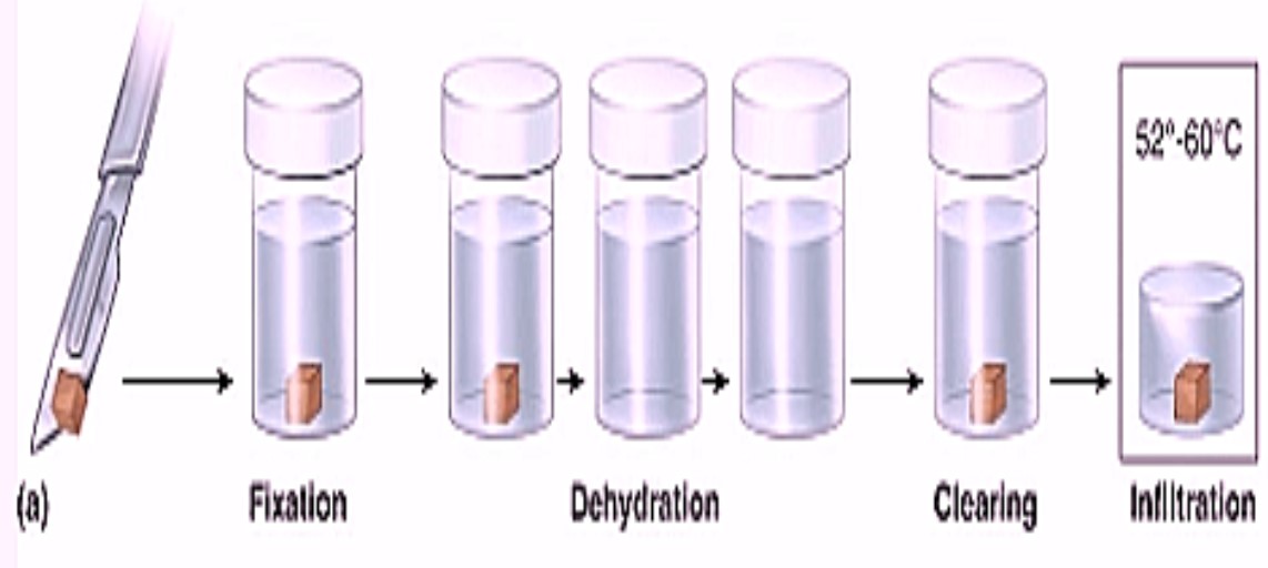
Fixation: Preserving the tissue’s structure by killing cells and preventing decomposition.
Dehydration: Removing water from the tissue to prepare it for embedding.
Embedding: Infiltrating the tissue with a solid medium (e.g., paraffin, resin) for support during sectioning.
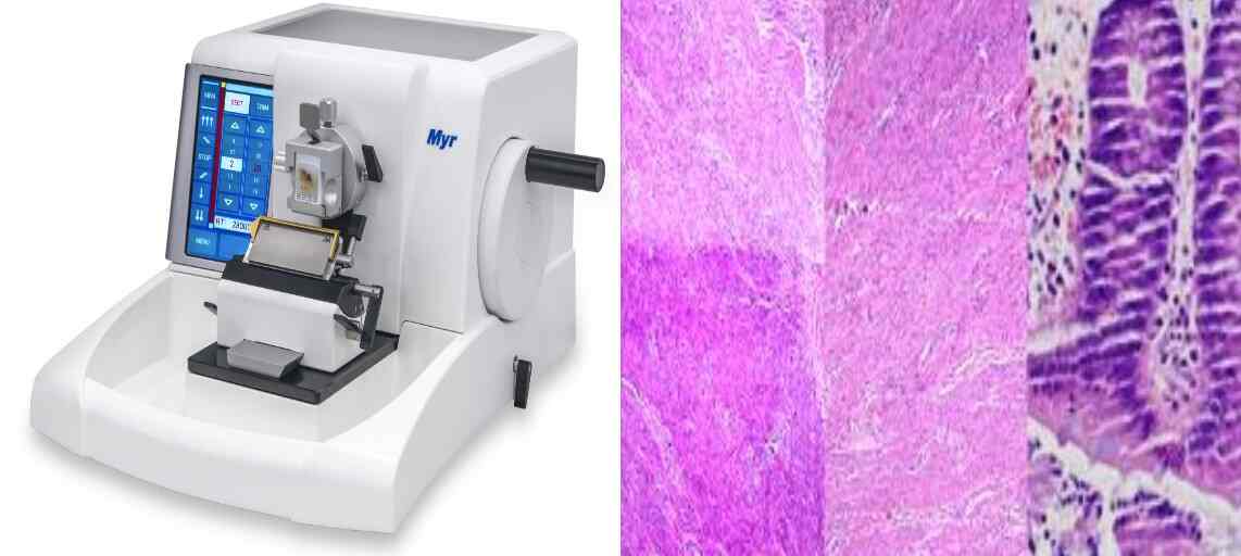
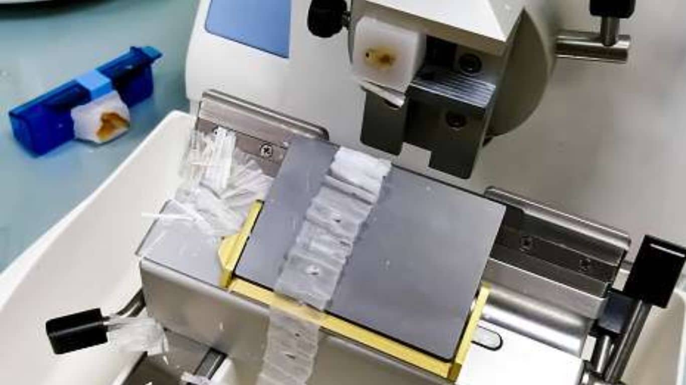
Sectioning: Cutting the embedded tissue into thin slices for microscopic examination.
Staining: Coloring the tissue to enhance contrast and visibility of specific structures.
Mounting: Placing the stained sections on a slide and covering them with a protective layer.
Advancements in Microtechnique
The field of microtechnique has seen significant advancements, driven by technological innovations and interdisciplinary research.
1. Electron Microscopy: Electron microscopy offers unparalleled resolution, allowing scientists to visualize structures at the nanometer scale. Techniques like transmission electron microscopy (TEM) and scanning electron microscopy (SEM) have revolutionized our understanding of cellular and material ultrastructure.
2. Confocal Microscopy: Confocal microscopy provides high-resolution, three-dimensional images of specimens by eliminating out-of-focus light. This technique is widely used in cell biology, neuroscience, and developmental biology for detailed imaging of complex structures.
3. Digital Imaging and Analysis: Advances in digital imaging and analysis have enhanced the capabilities of microtechnique. Automated image analysis software enables the quantification of microscopic features, improving the accuracy and efficiency of data interpretation.
4. Fluorescence Microscopy: Fluorescence microscopy utilizes fluorescent dyes and proteins to label specific cellular components, allowing for the visualization of dynamic processes within living cells. Techniques like super-resolution microscopy have pushed the boundaries of spatial resolution.
5. Cryo-Techniques: Cryo-techniques, such as cryo-electron microscopy (cryo-EM), involve freezing specimens to preserve their native state. Cryo-EM has been instrumental in elucidating the structures of macromolecular complexes at near-atomic resolution.
Conclusion
Microtechnique stands as a cornerstone of modern scientific research, providing the tools and methods necessary to explore the microscopic world. Its applications in histology, cytology, microbiology, materials science, and forensic science underscore its versatility and importance. As technology continues to advance, microtechnique will undoubtedly play a pivotal role in shaping the future of science and medicine.