
Taenia solium: Taenia solium, commonly known as the pork tapeworm, is a parasitic flatworm that infects both humans and pigs. Understanding its morphology is crucial not only for the identification and diagnosis of infections but also for developing effective treatments and preventive measures. This article delves into the detailed morphological features of Taenia solium.
Its life history is completed in two hosts, i.e., digenetic; man being the primary host and pig as secondary host. Except the adult, various stages of its life history are passed in the body of secondary host. Other animals like goat, cattle, horse and monkey may also serve as secondary host.
What is Taenia solium?
Taenia solium belongs to the class Cestoda, which includes all tapeworms. It is a zoonotic parasite, meaning it can be transmitted from animals to humans, typically through the ingestion of undercooked pork contaminated with larval cysts. The adult tapeworm lives in the human intestine, where it can grow to several meters in length.
Morphological Features of Taenia solium
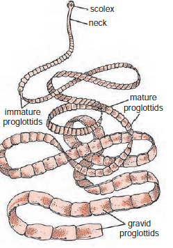
The morphology of Taenia solium is both fascinating and highly adapted to its parasitic lifestyle. The worm’s body is divided into three distinct regions: the scolex (head), the neck, and the strobila (body). Each of these regions plays a vital role in the worm’s survival and reproduction.
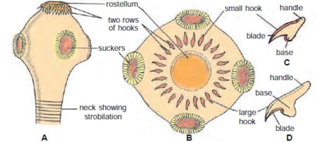
The Scolex
The scolex is the anterior (front) part of the tapeworm and is primarily responsible for attachment to the host’s intestinal wall. It is a small, bulbous structure equipped with both suckers and hooks.
The scolex is smaller than the head of a pin about 1 mm in diameter with 4 cup-like muscular suckers having radial muscles, and an anterior round prominence, the rostellum having about 22 to 32 curved, chitinous hooks in two circles, the inner circle with larger hooks and outer circle with smaller ones. The long and short hooks alternate with each other. Each hook is made of a base by which it fixes, a handle directed towards the apex and a conical blade directed outwardly. The rostellum can be protruded slightly. The scolex with its suckers and hooks is an organ of attachment to the intestinal wall of the host.
The Neck Region
The neck is a short, unsegmented region located just behind the scolex. This area is crucial for the growth and development of the tapeworm. The neck is where new segments, or proglottids, are formed. It acts as a growth zone, continually producing new segments as the worm matures and lengthens. As the neck generates new proglottids, these segments gradually mature and move down the strobila.
The Strobila
The strobila, or the main body of the tapeworm, is composed of a series of segments called proglottids. These segments are arranged in a chain-like fashion and represent different stages of development.
The strobila can grow to impressive lengths, sometimes reaching up to 7 meters in adult worms. The number of proglottids can range from hundreds to thousands, depending on the worm’s age and size. Proglottids play a critical role in the worm’s life cycle, with each segment housing a complete set of reproductive organs.
Immature Proglottids
The proglottids closest to the neck are immature and do not yet contain fully developed reproductive organs. These include nearly 200 anterior proglottids. These segments are smaller and less complex, primarily functioning in nutrient absorption and growth.
Mature Proglottids
As proglottids move further down the strobila, they reach maturity. At this stage, they contain fully developed male and female reproductive organs. Nearly 100 to 150 proglottids, after immature proglottids, bear male reproductive organs only. As these proglottids are pushed back they develop female reproductive organs also. Thus, the mature proglottids are hermaphrodite and they number about 300 to 400 proglottids.

Gravid Proglottids
The proglottids at the posterior end of the strobila are gravid, meaning they are filled with fertilized eggs. These proglottids are the oldest and towards the posterior side of the strobila and include nearly 150 to 200 proglottids. These segments are longer than their breadth and no reproductive organs are found in them. They contain only branched uterus packed with fertilised eggs.
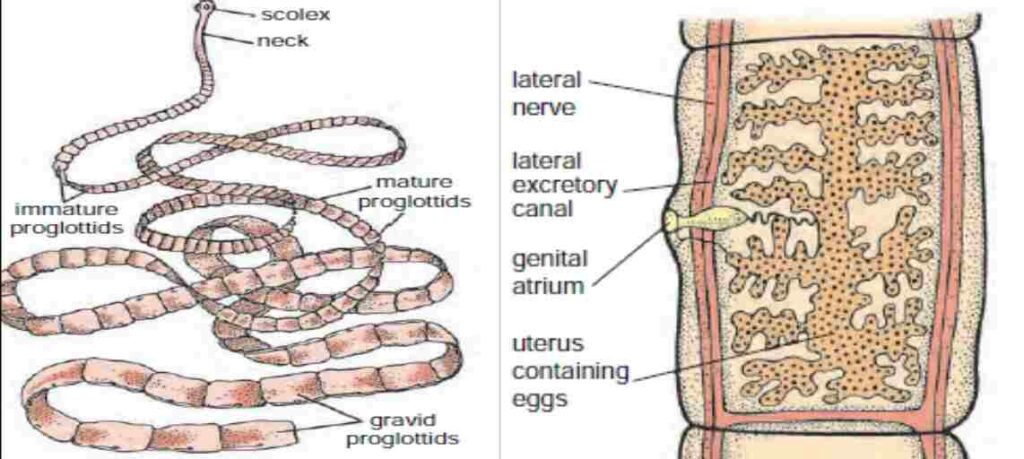
Gravid proglottids are larger and more elongated than their immature and mature counterparts. They are essentially egg sacs, ready to detach from the strobila. These segments eventually break off and are excreted with the host’s feces, spreading the eggs into the environment and continuing the parasite’s life cycle.
Tegument of Taenia solium
The tegument, or outer covering, of Taenia solium is a specialized structure that plays multiple roles in the worm’s survival. The tegument is a syncytial layer (a multinucleated mass without distinct cell boundaries) that covers the entire body. It facilitates the absorption of nutrients and helps protect the worm from the host’s immune system.
The surface of the tegument is covered with microtriches, tiny hair-like structures that increase the surface area for absorption and enhance the worm’s ability to adhere to the host’s intestinal walls.