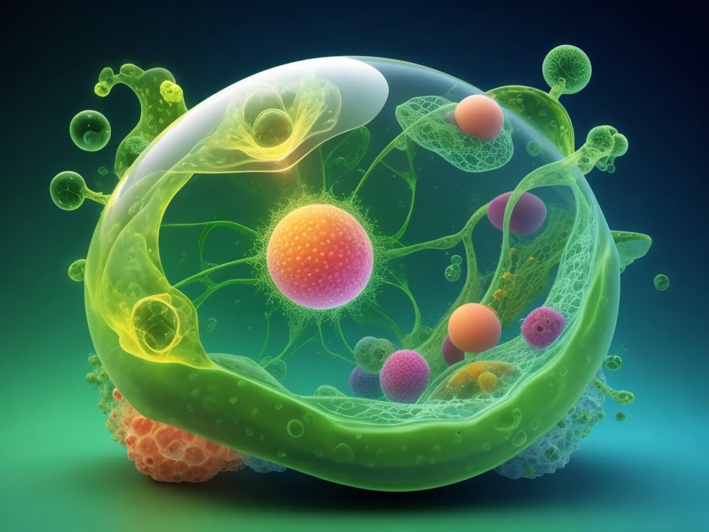


Ribosomes: Ribosomes consists of two units one small and other large which fit together and work as one to translate mRNA into a polypeptide chain during protein synthesis.
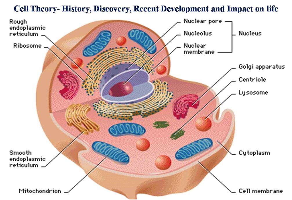
In prokaryotic cells ribosomes are synthesized in the cytoplasm and nucleolus in eukaryotic cells. Over 200 proteins are needed to synthesize the four types of RNA. Ribosomes can be found freely suspended in the cytoplasm or they are bound to the Endoplasmic reticulum or nuclear envelope.
Ribosomes are composed of 65% ribosomal RNA and 35% ribosomal proteins known as ribonucleoproteins. Ribosomes translates mRNA using amino acids of tRNA to build a polypeptide chain or proteins.
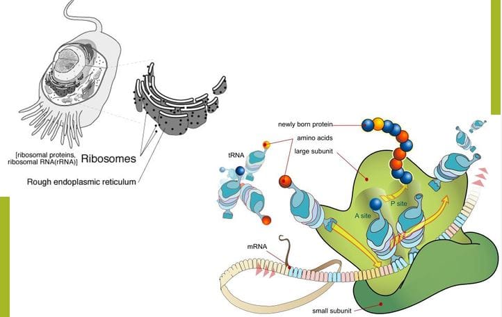
The newly formed proteins or polypeptide chains are inserted directly into ER by the ribosomes and then transferred to their destination. Mitochondria consist of ribosomes same as that of bacteria but they are not affected because of the double membrane which does not allow the antibiotics to come in the organelle.
Polyribosome /polysome is a cluster of ribosomes a single strand of messenger ribonucleic acid (mRNA) engaged in the synthesis of protein. The ribosomal subunits and their RNAs are named on the sedimentation rate unit, called as Svedberg unit or S value.
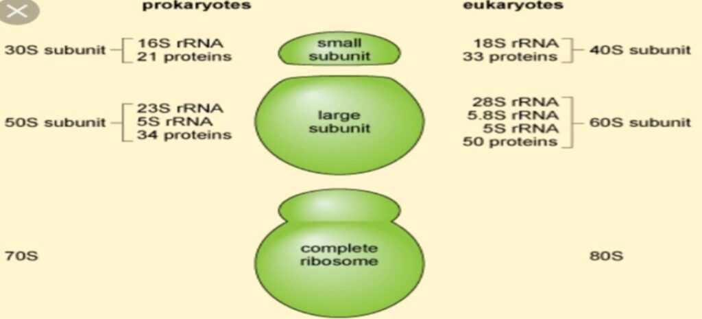
The difference between prokaryotic and eukaryotic cell ribosomes are exploited by the chemist of pharmaceuticals to produce antibiotics, that can destroy bacteria without harming the cells of infected persons. Due to the antibiotics the bacterial ribosomes of 70S are vulnerable but 80S of eukaryotes are not. Prokaryotes have 70S ribosomes each consisting 30S small and 50S large ribosomal subunit. Eukaryotes have 80S large and 40S small subunit. Eukaryotic ribosomes: 80S unit, having 60S and 40S subunits in it. Prokaryotic ribosomes are 70S, having 50S and 30S subunits.
| Robosomes | Subunits | rRNA | Proteins |
| 80s (Eukaryotic) | 60S | 5.8S, 5S, 28S | ~49 |
| 40S | 18S | ~33 | |
| 70s (Prokaryotic) | 50S | 23S, 5S | 34 Proteins L1-L34 |
| 30S | 16S | 21 Proteins S1-S21 |
Types of Ribosomes:
Ribosomes are essential molecular machines found in all living cells, and they come in two main types based on their location and structural differences:
1. Prokaryotic Ribosomes (70S)
- Found in: Bacteria and Archaea
- Size: 70S (Svedberg units, which reflect sedimentation rate, not mass)
- Subunits:
- Large subunit (50S): Contains 23S rRNA, 5S rRNA, and proteins
- Small subunit (30S): Contains 16S rRNA and proteins
Smaller and less complex than eukaryotic ribosomes
Targeted by many antibiotics (e.g., tetracycline, streptomycin), which do not affect eukaryotic ribosomes due to structural differences
2. Eukaryotic Ribosomes (80S)
- Found in: Animal cells, plant cells, fungi, protists
- Size: 80S
- Subunits:
- Large subunit (60S): Contains 28S rRNA, 5.8S rRNA, 5S rRNA, and proteins
- Small subunit (40S): Contains 18S rRNA and proteins
Larger and more complex, Located in two main regions:
- Free ribosomes (floating in the cytoplasm): Synthesize proteins for use inside the cell
- Bound ribosomes (attached to rough endoplasmic reticulum): Make proteins for secretion or membrane insertion
3. Organelle Ribosomes (70S-like)
Found in: Mitochondria (in all eukaryotes), Chloroplasts (in plants and algae)
- Size: 70S (similar to prokaryotic ribosomes)
- Subunits: Large (50S) and Small (30S)
Involved in synthesis of a small number of organelle-specific proteins. Resemble prokaryotic ribosomes in structure and function. Support the endosymbiotic theory (mitochondria and chloroplasts originated from prokaryotic ancestors)
Structure of Ribosomes:
Ribosomes are essential cellular machinery responsible for protein synthesis (translation). They are complex molecular structures composed of ribosomal RNA (rRNA) and ribosomal proteins (r-proteins). While their fundamental function is conserved across all life forms, their exact composition and size differ between prokaryotic and eukaryotic cells.
I. General Features:
Ribosomes are made of both RNA and protein, making them a ribonucleoprotein complex. Every ribosome consists of two distinct subunits:
- Small Subunit: This subunit is responsible for binding and decoding the messenger RNA (mRNA). It reads the genetic code carried by the mRNA.
- Large Subunit: This subunit is where amino acids are joined together to form the polypeptide chain. It contains the peptidyl transferase center (PTC), which catalyzes the formation of peptide bonds between amino acids.
- Svedberg Units (S): Ribosomal subunits are characterized by their sedimentation rate in a centrifuge, measured in Svedberg units (S). This unit reflects their size, shape, and density. It’s important to note that the Svedberg units of the subunits don’t simply add up to the total Svedberg unit of the complete ribosome (e.g., 30S + 50S = 70S, not 80S).
- Binding Sites: Both the small and large subunits come together to form a cleft or channel through which the mRNA passes. Within this structure, there are three key binding sites for transfer RNA (tRNA) molecules:
- A (Aminoacyl) site: Accommodates the incoming tRNA carrying a new amino acid.
- P (Peptidyl) site: Holds the tRNA with the growing polypeptide chain.
- E (Exit) site: Where the deacylated (empty) tRNA exits the ribosome.
Prokaryotic Ribosomes (70S):
Prokaryotic ribosomes (found in bacteria and archaea) are smaller and less complex than eukaryotic ribosomes.
- Total Size: 70S
- Subunits:
- Small Subunit (30S): Composed of one 16S rRNA molecule and approximately 21 ribosomal proteins. The 16S rRNA plays a crucial role in mRNA binding and decoding.
- Large Subunit (50S): Composed of two rRNA molecules (23S rRNA and 5S rRNA) and approximately 31 ribosomal proteins. The 23S rRNA contains the peptidyl transferase activity, meaning it’s the catalytic core of the ribosome.
Eukaryotic Ribosomes (80S):
Eukaryotic ribosomes are larger and more complex.
- Total Size: 80S
- Subunits:
- Small Subunit (40S): Composed of one 18S rRNA molecule and about 33 ribosomal proteins. The 18S rRNA is important for recognizing the start codon on the mRNA.
- Large Subunit (60S): Composed of three rRNA molecules (28S rRNA, 5.8S rRNA, and 5S rRNA) and approximately 49 ribosomal proteins. Like in prokaryotes, the large rRNA molecule (28S rRNA) in eukaryotes is responsible for peptidyl transferase activity.
Location of Ribosomes: Ribosomes can be found in various locations within the cell:
- Free ribosomes: Suspended in the cytoplasm, they synthesize proteins destined for use within the cell.
- Membrane-bound ribosomes: Attached to the endoplasmic reticulum (forming the rough ER) or the nuclear envelope. These ribosomes synthesize proteins that are destined for secretion outside the cell, insertion into membranes, or delivery to organelles like lysosomes.
- Mitochondrial and Chloroplast ribosomes: These organelles within eukaryotic cells also contain their own ribosomes, which are structurally similar to prokaryotic 70S ribosomes, reflecting their endosymbiotic origin.
How does the ribosomal movement take place in translation?
During translation, ribosomal movement is a highly coordinated process that ensures accurate synthesis of proteins. The ribosome moves along the messenger RNA (mRNA) in a specific direction (5′ to 3′), decoding it and building the corresponding polypeptide chain. Here’s how ribosomal movement takes place:
1. Initiation (Positioning the Ribosome)
- Translation begins at the start codon (AUG) on the mRNA.
- The small ribosomal subunit binds to the mRNA near the 5′ cap (in eukaryotes) or Shine-Dalgarno sequence (in prokaryotes).
- The initiator tRNA carrying methionine binds to the start codon.
- The large ribosomal subunit then joins to form the complete ribosome.
2. Elongation (Main Movement Phase)
This is where the ribosome actively moves along the mRNA, decoding codons and forming the polypeptide chain.
The ribosome has 3 sites:
- A site (Aminoacyl-tRNA site) – entry point for new tRNAs.
- P site (Peptidyl-tRNA site) – holds the tRNA with the growing peptide chain.
- E site (Exit site) – where empty tRNAs exit.
Each elongation cycle involves:
- Codon Recognition:
- A charged tRNA enters the A site, matching the codon on mRNA.
- Peptide Bond Formation:
- The growing peptide is transferred from the tRNA in the P site to the new amino acid on the tRNA in the A site.
- This reaction is catalyzed by peptidyl transferase activity of the ribosome.
- Translocation:
- The ribosome shifts one codon downstream (5′ → 3′ direction on mRNA).
- The tRNA that was in the P site moves to the E site and exits.
- The tRNA with the growing chain moves from the A site to the P site.
- The A site becomes empty again for the next incoming tRNA.
This cycle repeats until a stop codon is reached.
3. Termination (Ending Translation)
Disassemble into subunits, freeing the mRNA.
When a stop codon (UAA, UAG, or UGA) enters the A site, no matching tRNA binds.
Instead, release factors bind and cause the ribosome to:
Release the finished polypeptide.
Function:
Ribosomes are essential for protein synthesis, acting as the sites where genetic instructions are translated into functional proteins. Here are the key functions of ribosomes:
1. Protein Synthesis (Translation)
- Primary function of ribosomes.
- They read the sequence of codons on messenger RNA (mRNA) and match them with the appropriate amino acids.
- This process has three major steps:
- Initiation: Ribosome assembles on the start codon of mRNA.
- Elongation: Ribosome moves along mRNA, adding amino acids to the growing polypeptide chain.
- Termination: Ribosome stops at a stop codon and releases the completed protein.
2. Decoding Genetic Information
- Ribosomes read mRNA codons and ensure that the correct transfer RNA (tRNA) carrying a specific amino acid pairs with the mRNA.
- This guarantees that the genetic code is accurately translated into an amino acid sequence.
3. Peptide Bond Formation
- Ribosomes catalyze the formation of peptide bonds between adjacent amino acids during translation.
- This enzymatic activity is carried out by the peptidyl transferase center located in the large ribosomal subunit.
4. Quality Control of Translation
- Ribosomes help ensure fidelity and accuracy by:
- Checking tRNA-mRNA base pairing
- Using elongation factors and proofreading steps to reduce errors
- Recognizing premature stop codons or damaged mRNA (with help from other quality control systems)
5. Location-Dependent Functions
- Free Ribosomes:
- Synthesize proteins that remain in the cytosol or go to organelles like the nucleus, mitochondria, or peroxisomes.
- Bound Ribosomes (on rough ER):
- Synthesize secretory, membrane-bound, or lysosomal proteins.
- These proteins are typically exported from the cell or inserted into membranes.
6. Supporting Organelle Protein Production
Ribosomes in mitochondria and chloroplasts produce a small set of organelle-specific proteins, essential for the function of those organelles.