Skip to content
Zoology Library Logo

Zoology Library

Zoology Library

  • Home
  • Biology
    • Zoology
    • Developmental Biology
    • SEC/VSC Cources
      • SEC Course on Microtechnique
      • Skill Enhancement Course on Haematology
      • Skill Enhancement Course on Urinology
      • SEC on Aquarium Fish Keeping
  • Economic Zoology
  • Fishery Science
  • Cell Biology
  • Conservation
  • Zoo Park & News
  • Web Stories
  • Toggle search form
  • Brackishwater Fishes: Characters and Classification Fishery Science
  • Kolleru Lake, India
    Kolleru Lake: Fisheries Fishery Science
  • Sewage Fed Fisheries in India Fishery Science
  • Importance of nematodes for Crop production
    Importance of Nematodes for Crop Production Economic Zoology
  • Bee Keeping Methods and Equipments Economic Zoology
  • Phylum Cnidaria
    Phylum Cnidaria Characteristic and Classification Zoology
  • Cleavage Stages of Embryonic Development
    Cleavage Stages of Embryonic Development Developmental Biology
  • Species suitable for Biofloc Fish Farming in India
    Species suitable for Biofloc Fish Farming in India Fishery Science
Protozoa slides

Protozoa: Slides from Ciliates, Opalinates, and Flagellates

Posted on November 11, 2023August 7, 2024 By Rajkumar Pawar No Comments on Protozoa: Slides from Ciliates, Opalinates, and Flagellates

Protozoa: Protozoans are single-celled eukaryotic organisms that are found in a variety of habitats, including water, soil, and the bodies of animals. They can be classified into four main groups based on their locomotion: flagellates, ciliates, amoebae, and sporozoans.

The slides are used to study the morphology of protozoans under a microscope. The slides are usually prepared by staining the protozoans with a dye, such as methylene blue or carmine. This makes the single cell animal more visible under the microscope and allows you to see their different structures.

There are many different types of protozoans, and they can be found in a variety of habitats, including water, soil, and the bodies of animals. Some protozoans are harmless to humans, while others can cause diseases. Protozoan slides can be used to identify different types of protozoans, to study their morphology, and to investigate their behavior. They can also be used to diagnose diseases caused by protozoans.

Protozoa: Slides from Ciliates, Opalinates, and Flagellates

Some common types of protozoan, slides include:

1. Opalina Purkinye & Valentin, 1835
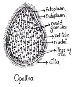
Protozoa- Oplania

Classification

  • Kingdom: Chromista
  • Subkingdom: Protista
  • Phylum: Sarcomastigophora
  • Subphylum: Sarcodina
  • Class: Opalinea
  • Order: Opalinida
  • Family: Opalinidae
  • Genus: Opalina

Comments:

  1. Commonly referred as rectal ciliate
  2. Body is simple oval and flattened
  3. Measures upto 400-500µ in diameter
  4. Whole body is covered with cilia
  5. Opalina is multinucleated.
  6. Found in rectum of Amphibians

2. Nyctotherus
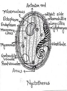
Protozoa-Nyctotherus

  • Kingdom: Protista
  • Subikingdom: Baciliata
  • Phylum: Ciliophora
  • Subphylum: Intramacronucleata
  • Class: Spirotrichea
  • Order: Clevelandellida
  • Family: Nyctotheridae
  • Genus: Nyctotherus

Comments:

  1. Found in alimentary canal of cockroaches, fishes, amphibians and mammals
  2. Body bean shaped and dorsoventrally flattened
  3. Measures upto 60-120 µ in diameter
  4. Clear distinction between ectoplasm and endoplasm
  5. Two nuclei are present, micro and macronucleus
  6. Body covered with cilia.

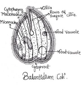
3. Balantidium coli

Classification

  • Kingdom: ProtistaProtozoa - Balantidium-coli
  • Phylum: Ciliophora
  • Class: Litostomatea
  • Order: Vestibuliferida
  • Family: Balantidiidae
  • Genus: Balantidium
  • Species: coli

Comments:

  1. Commonly found in the intestine of pigs, sheep, camels, ostrich, cockroaches and man
  2. Spherical in shape and motile
  3. Measures 60-70 µ in diameter
  4. Body covered with spiral rows of cilia
  5. Endoplasm contains micronucleus and sausage shaped macronucleus.

4. Trichomonas
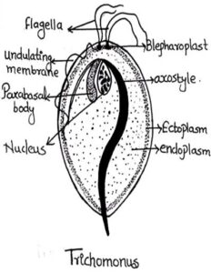
Protozoa -Trichomonas

Classification

  • Kingdom: Protista
  • Phylum: Metamonada
  • Class: Parabasillia
  • Order: Trichomonadida
  • Family: Trichomonadidae
  • Genus: Trichomonas
  • Species: vaginalis

Comments:

  1. Found in intestine of vertebrates, leeches and termites and in man it is found in vagina, colon and buccal cavity
  2. Body oval, egg shaped slightly tapering posteriorly
  3. 5-20 µ in length
  4. 3-5 flagella are present
  5. Flagellum arising from the left blepharoplast forms the undulating membrane.

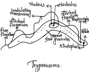
5. Trypanosoma

Classification

  • Protozoa - TrypanosomaKingdom: Protista
  • Phylum: Sarcomastigophora
  • Class: Zoomastigophorea
  • Order: Trypanosomatida
  • Family: Trypanosomatidae
  • Genus: Trypanosoma


Comments:

  1. It is the blood parasite of man
  2. Body elongated, leaf like, flattened and tapering at both the ends
  3. Measures 10-40 µ in length and 2-10 µ in breadth
  4. Single flagellum running along the edge of undulating membrane
  5. Large central nucleus with nucleolus is present.
Zoology Tags:Zoology

Post navigation

Previous Post: Discovering the Charm of Indira Gandhi Zoological Park in Visakhapatnam, Andhra Pradesh
Next Post: National Zoological Park, New Delhi

Related Posts

  • Phylum Platyhelminthes Characters, Classification, Examples and Recent Discovery Zoology
  • Coelomate Animals: Museum Specimens Zoology
  • Metamorphosis in Insects (Arthropods) Zoology
  • Phylum Echinodermata–General Characters and Classification upto Classes level Zoology
  • Aschelminthes/Nemathelminthes Characteristic and Classification Zoology
  • Recent Trends, Scope and Career in Zoology Zoology

Leave a Reply Cancel reply

Your email address will not be published. Required fields are marked *

  • Exotic and Endemic species of Aquarium fishes
    Exotic and Endemic species of Aquarium fishes SEC/VSC Courses
  • Captivating Wildlife at Indira Gandhi Zoological Park
    Discovering the Charm of Indira Gandhi Zoological Park in Visakhapatnam, Andhra Pradesh Zoo Park & News
  • Exploitation of Reservoir Fisheries in India Fishery Science
  • Ghost Frog
    Ghost Frog Rediscovered After 130 Years

    Ghost Frog: Researchers meticulously studied Germain’s travel accounts and other historical documents from the late 19th century. By reconstructing his likely route through the estate, they identified probable collection sites in the remote Andean foothills Ghost Frog Species: Alsodes vittatusLast Seen: 1902Rediscovery: 2024, Chile Species Last Seen Rediscovered Location Habitat Type Conservation Status Alsodes vittatus 1902 2024 La...<p class="more-link-wrap"><a href="https://zoolibs.com/ghost-frog-rediscovered-after-130-years/" class="more-link">Read More<span class="screen-reader-text"> “Ghost Frog Rediscovered After 130 Years”</span> »</a></p>

  • vermiculture
    Vermiculture Economic Zoology
  • Induced Breeding Technique of fishes (Hypophysation) Fishery Science
  • Value Added Fish Products Fishery Science
  • Microtechnique: Definition, Scope and Application SEC Course on Microtechnique

Categories

  • Biology
  • Cell Biology
  • Conservation
  • Developmental Biology
  • Economic Zoology
  • Fishery Science
  • SEC Course on Microtechnique
  • SEC on Aquarium Fish Keeping
  • SEC/VSC Courses
  • Skill Enhancement course on Haematology
  • Skill Enhancement Course on Urinology
  • Zoo Park & News
  • Zoology

Recent Posts

  • Scoliodon (Dogfish)
  • New Change Coming in Higher Education!!!
  • Torsion in Gastropods
  • Ribosomes: Structure and Functions
  • An Overview of Fisheries Development in India

Pages

  • About Us
  • Contact Us
  • Disclaimer
  • Privacy Policy
  • Terms and Conditions

Copyright © 2026 Zoology Library.

Powered by PressBook News WordPress theme