Cleavage Stages of Embryonic Development: Embryonic development is a complex and highly regulated process that begins with the fertilization of an egg by a sperm and continues through various stages to form a multicellular organism. One of the earliest and crucial processes in embryonic development is cleavage. Cleavage refers to a series of rapid cell divisions that occur shortly after fertilization. These divisions are essential for increasing the number of cells and establishing the basic body plan of the developing embryo. Cleavage is the process of rapid cell division that occurs in the early stages of embryonic development. It begins after the zygote, or fertilized egg, forms, and continues until the blastula is formed.
Salient Features of Cleavage
The salient features of cleavage are as follows:
- Cleavage is a rapid process of cell division that occurs in the early stages of embryonic development.
- It begins after the zygote, or fertilized egg, forms, and continues until the blastula is formed.
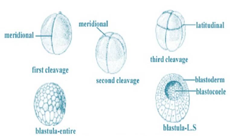
- The blastula is a hollow sphere of cells that surrounds a fluid-filled cavity called the blastocoel.
- The cells of the blastula are called blastomeres.
- There are two main types of cleavage: holoblastic cleavage and meroblastic cleavage.
- Cleavage is a mitotic process, meaning that the daughter cells are genetically identical to the parent cell.
- Cleavage does not increase the total volume of the embryo. Instead, the cytoplasm of the zygote is divided among the blastomeres.
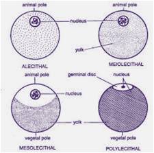
- Cleavage patterns are characteristic of different species. This is because cleavage patterns are influenced by the amount of yolk in the egg and the overall shape of the embryo.
- Cleavage is a critical stage in embryonic development because it allows the zygote to grow and develop into a multicellular embryo.
Types and patterns of Cleavage
There are two main types of cleavage: holoblastic cleavage and meroblastic cleavage.
Holoblastic cleavage
Holoblastic cleavage is a type of cleavage in which the entire zygote divides into smaller and smaller cells. Holoblastic cleavage is further divided into four subtypes: radial, spiral, bilateral, and rotational.
Radial cleavage is a type of holoblastic cleavage in which the cleavage planes divide the zygote into equal halves, then quarters, then eighths, and so on. Radial cleavage is common in echinoderms (such as sea stars and sea urchins) and amphibians.
Spiral cleavage is a type of holoblastic cleavage in which the cleavage planes divide the zygote in a spiral pattern. Spiral cleavage is common in annelids (such as earthworms and leeches), mollusks (such as snails and clams), and arthropods (such as insects and spiders).
Bilateral cleavage is a type of holoblastic cleavage in which the cleavage planes divide the zygote into two halves, then quarters, then eighths, and so on, but the halves are not identical. Bilateral cleavage is common in chordates (such as humans, fish, amphibians, reptiles, birds, and mammals).
Rotational cleavage is a type of holoblastic cleavage in which the cleavage planes rotate slightly after each division. Rotational cleavage is common in nematodes (such as roundworms) and some insects.
Meroblastic cleavage
Meroblastic cleavage is a type of cleavage in which only a portion of the zygote divides. This is because some zygotes have a large amount of yolk, which is a nutrient-rich material that provides food for the developing embryo. Meroblastic cleavage is further divided into two subtypes: discoidal and superficial.
Discoidal cleavage is a type of meroblastic cleavage in which the cytoplasm of the zygote is divided into a disc-shaped region at the top and a yolk-filled region at the bottom. Discoidal cleavage is common in reptiles and birds.
Superficial cleavage is a type of meroblastic cleavage in which the cytoplasm of the zygote is divided into a thin layer of cells at the top and a yolk-filled region at the bottom. Superficial cleavage is common in insects.
Cleavage is a critical stage in embryonic development because it allows the zygote to grow and develop into a multicellular embryo. The different types of cleavage patterns that occur in different species are thought to be related to the amount of yolk in the egg and the overall shape of the embryo.
Cleavage patterns are also important for determining the fate of different cells in the embryo. For example, in radial cleavage embryos, the cells at the top of the blastula eventually form the ectoderm, while the cells at the bottom of the blastula eventually form the endoderm.
Cleavage Stages in Humans
Human embryos undergo holoblastic cleavage. The first cleavage division occurs about 24 hours after fertilization, and the resulting two cells are called blastomeres. The blastomeres continue to divide rapidly, and by the fourth day of development, the embryo is a ball of 16 cells called the morula. The morula continues to divide, and by the fifth day of development, it has formed a hollow sphere of cells called the blastocyst. The blastocyst is made up of an outer layer of cells called the trophoblast and an inner cell mass. The inner cell mass will eventually develop into the embryo, while the trophoblast will form the placenta.
The blastocyst implants in the uterine wall on the sixth or seventh day of development. After implantation, the embryo continues to develop and grow, eventually forming a fetus.
Stages of Embryonic Development
Embryonic development is begins with the fertilization of an egg by a sperm and continues through various stages to form a multicellular organism, these stages are as follows.
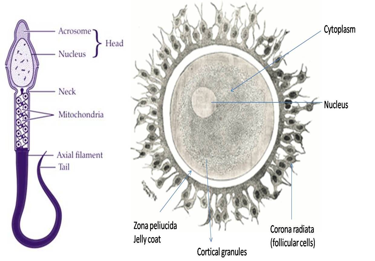
1. Fertilization: Fertilization is the initial step in embryonic development. It occurs when a sperm cell successfully penetrates the egg cell, resulting in the formation of a zygote. The zygote contains the combined genetic material from both the mother and the father, setting the stage for the development of a new organism.
2. First Cleavage: The first cleavage typically occurs shortly after fertilization and is often referred to as the two-cell stage. During this stage, the zygote undergoes its first division, giving rise to two daughter cells called blastomeres. These blastomeres are genetically identical and represent the earliest stage of embryonic development.
3. Second Cleavage: Following the first cleavage, the embryo progresses to the two-cell stage, where each of the two blastomeres divides again. This results in the formation of four blastomeres. At this point, the embryo is referred to as a four-cell embryo.
4. Morula Stage: As cleavage continues, the embryo undergoes further divisions, leading to the formation of a solid ball of cells known as the morula. The morula consists of multiple blastomeres and resembles a mulberry in appearance. During this stage, the embryo remains enclosed within the zona pellucida, a protective glycoprotein layer.
5. Blastula Stage: The next stage of development is the blastula stage. Here, the morula continues to divide, forming a hollow, fluid-filled structure called the blastocyst. The blastocyst consists of two main cell types: the inner cell mass (ICM), which will give rise to the embryo itself, and the outer layer of cells called the trophoblast, which will contribute to the formation of extraembryonic tissues.
6. Implantation: Following the blastula stage, the blastocyst undergoes implantation, which is the process by which it attaches to the uterine lining (endometrium). Successful implantation is crucial for the embryo to establish a connection with the maternal blood supply, which provides essential nutrients and support for further development.
7. Gastrulation: Gastrulation is a pivotal stage in embryonic development during which the three primary germ layers—ectoderm, mesoderm, and endoderm—are established. These germ layers give rise to various tissues and organs in the developing organism. Gastrulation involves complex cellular movements and rearrangements.
8. Neurulation: Neurulation is a specific process within embryonic development where the neural tube, the precursor to the central nervous system (brain and spinal cord), forms from the ectoderm. It is a critical step in the development of the nervous system.
9. Organogenesis: Organogenesis follows gastrulation and involves the differentiation and development of specific organs and tissues from the three germ layers. This stage is characterized by the formation of structures such as the heart, lungs, liver, and limbs, each with its unique developmental pathway.
Conclusion
The cleavage stages of embryonic development are a series of rapid cell divisions that set the foundation for the formation of a multicellular organism. These stages, from fertilization to organogenesis, are tightly regulated and intricately coordinated, leading to the remarkable process of embryogenesis. Understanding these stages is essential for researchers, healthcare professionals, and anyone interested in the fascinating world of embryonic development.